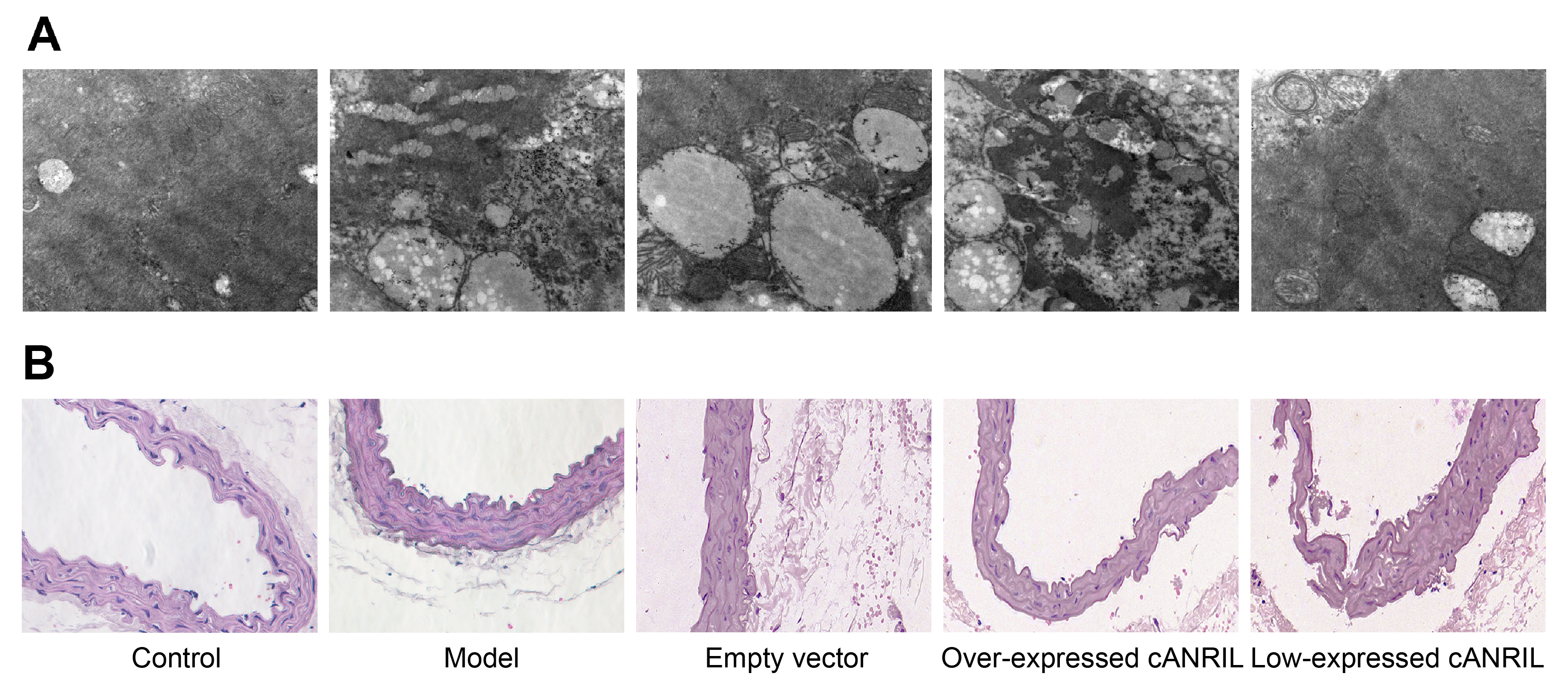
Fig. 2.Observation of ultrastructural and endothelial morphology of coronary arteries of rats in each group. Note: (A) shows a view of the ultrastructure of vascular ECs and SMCs from coronary arteries of rats in each group using an electron microscope (×10000), and (B) shows a view of the change in the endothelial mor¬phology of coronary arteries of rats in each group observed using HE staining (×200). EC, endothelial cell; SMC, smooth muscle cell; and cANRIL, circular antisense non-coding RNA in the INK4 locus.